 KOREAN
Protein Expression Analysis
Protein Expression Analysis
APOPTOSIS
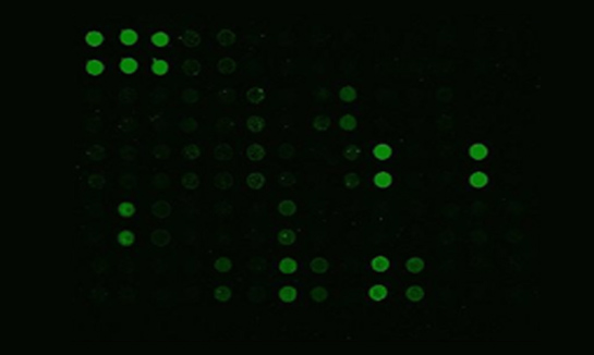
Apoptosis is a process of programmed cell death involving a series of biochemical events leading to characteristic cell morphology and death. Apoptosis is mediated by a diverse range of extracellular or intracellular cellular signals. After TNF and Fas activation, a balance is established between pro-apoptotic (BAX, BID, BAK or BAD) and anti-apoptotic (Bcl-X1 and Bcl-2) members of the Bcl-2 family. Using an antibody array kit, researchers can simultaneously detect the relative levels of 43 apoptosis-related proteins in cell lysates.
|
01
HUMAN APOPTOSIS ARRAY G1
It detects 43 Human Apoptotic Factors.
It is suitable for all liquid sample types, but please use with cell and tissue lysates.
G-Series Human Apoptosis Antibody Array 1Kit
| bad | bax | bcl-2 | bcl-w | BID |
| BIM | Caspase-3 | Caspase-8 | CD40 (TNFRSF5) |
CD40 Ligand (TNFSF5) |
| cIAP-2 | Cytochrome C | DR6 (TNFRSF21) |
Fas (TNFRSF6/Apo-1) |
Fas Ligand (TNFSF6) |
| HSP27 | HSP60 | HSP70 | HTRA2 | IGF-1 |
| IGF-2 | IGFBP-1 | IGFBP-2 | IGFBP-3 | IGFBP-4 |
| IGFBP-5 | IGFBP-6 | IGF-1 R | livin | p21 |
| p27 | p53 | SMAC | Survivin (BIRC5) |
TNF RI (TNFRSF1A) |
| TNF RII (TNFRSF1B) |
TNF alpha | TNF beta (TNFSF1B) |
TRAIL R1 (TNFRSF10A/DR4) |
TRAIL R2 (TNFRSF10B/DR5) |
| TRAIL R3 (TNFRSF10C) |
TRAIL R4 (TNFRSF10D) |
XIAP |
Research Service Implementation Process

STEP 1.
Study Design & NDA Writing

STEP 2. Microarray
Preparation &
Sample Pretreatment

STEP 3.
Main Experiment & Analysis

STEP 4.
Writing & Sending Report


